

The problem that won’t stay quiet
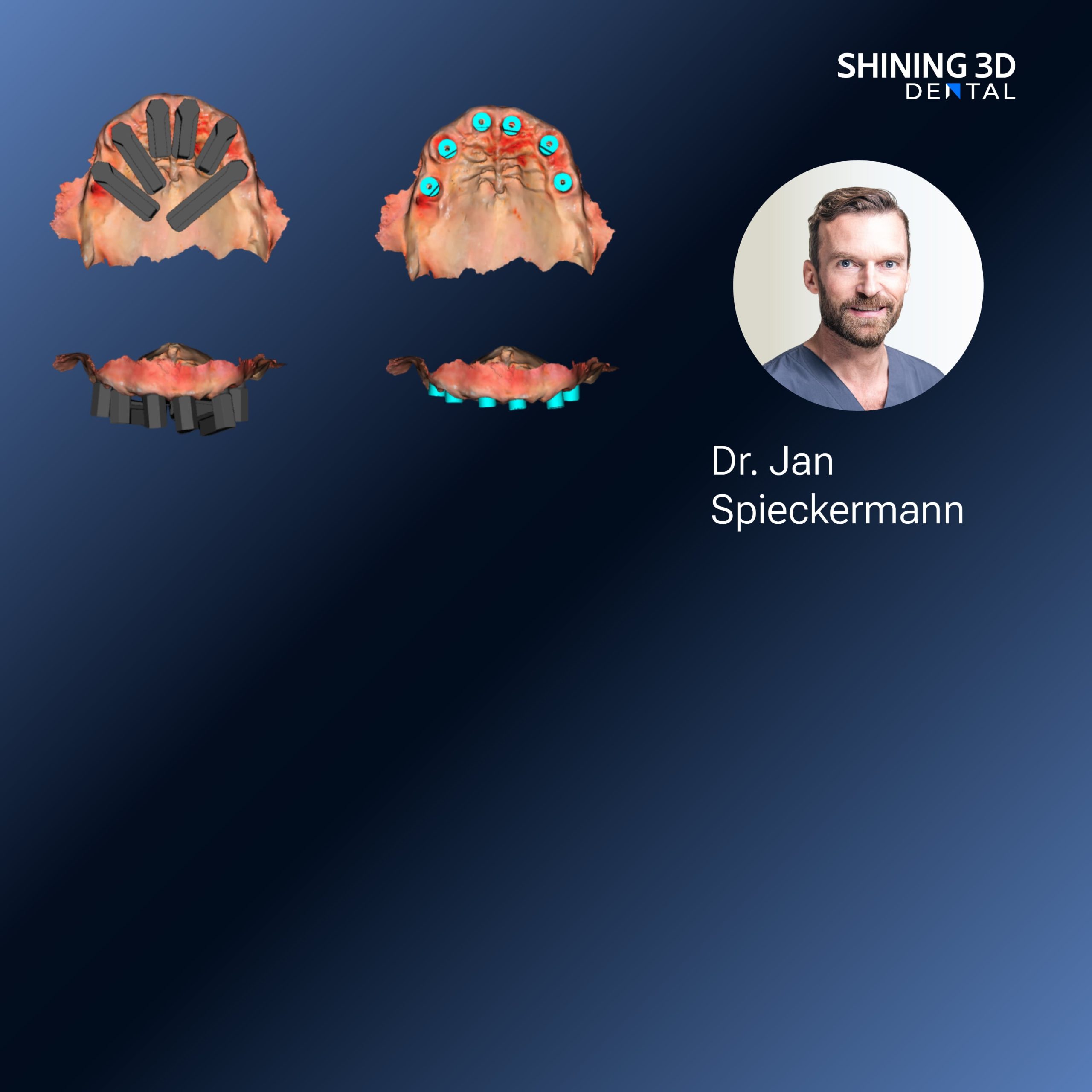
Clinics buy shiny boxes and promise patients a “digital” turnaround, but reality trips over fit issues and delayed lab feedback; the friction usually starts right after the first capture — the intraoral scanner image looks clean on screen but the crown that arrives doesn’t seat properly. That gap between a pretty file and a reliable prosthesis costs chair time, reputation, and calm nerves. Let’s treat this like a solvable engineering problem, not a mystery.
What’s actually causing the failures
Errors come from predictable places: incomplete scans, bad bite records, miscommunicated scanbody positions, and hidden software settings that change mesh resolution or file export names. Human factors — rushed scans, inconsistent angulation, or forgetting to capture opposing or buccal bite — amplify those technical flaws. The tools are only as good as the workflow around them.
A concise, problem-driven workflow that works
Start with intent: know the restoration type and required margins before you scan. Capture full arch or quadrant with deliberate overlap, then capture bite in a closed, repeatable posture. Verify scanbodies visually and retake if any interface looks noisy. Export open STL or PLY when the lab prefers freedom; use native formats only when you trust the ecosystem. Share a clearly labeled reference photo and a short note about occlusion and cementation strategy — small context removes big guesswork.
Common pitfalls and the small fixes that matter
Truncated margins: rescan with a slightly wider sweep and pull soft tissue with a dry tip. Misplaced scanbodies: confirm threads are seated and photograph indexed position. No occlusion: ask the patient to bite on a precise jig during capture. Mesh wobble from shaky hands: use short, overlapping passes and let the software stitch rather than forcing long sweeps. These fixes are low effort and high impact.
Alternatives worth weighing — concise trade-offs
Traditional impression still wins where tissue management is brutal or implant angulation is extreme, but it adds lab steps. Desktop scanners reduce clinic variability but add turnaround time for shipping. Closed, manufacturer-locked systems can deliver tight integration; open workflows give flexibility and better pricing from labs. Powder-free handhelds are faster and more patient-friendly; powdered systems sometimes produce crisper surface detail. Pick the compromise that matches your case mix and staff skills.

Real-world anchor and what it taught me
At the International Dental Show (IDS) in Cologne, clinicians and lab technicians demoed hands-on cases and repeatedly pointed to one recurring truth: seamless results hinge on repeatable capture and clear data handoff — a lesson visible across booths demonstrating the latest dental intraoral scanner technologies. Having observed those demonstrations and later worked with labs applying the same scan principles, I’ve seen marginal gaps drop and remakes halve when teams agreed on a few simple capture-and-communication rules.
Synthesis — a practical path forward
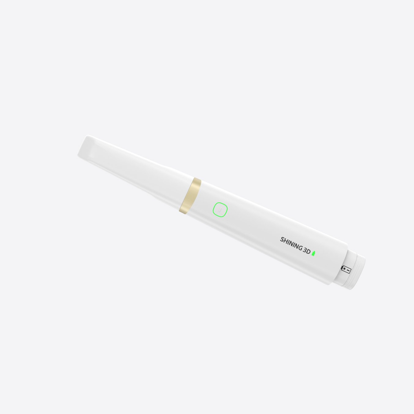
Fix the weak links: disciplined scanning, explicit lab notes, and choosing the right downstream process (milling or printing) for the material. When those elements line up, the digital promise moves from marketing-speak to routine. For teams building that continuity, tools and support from vendors and lab partners matter — and aligning with practical solutions like those from SHINING 3D DENTAL often makes the difference between another remake and a prosthesis that simply fits.